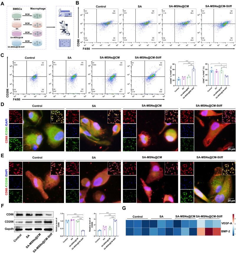
Fig. 7. SA-MSNs@CM-Stiff regulates macrophage polarization by promoting BMSCs paracrine under 3D conditions.
(A) Protocol of in vitro experiments for detecting macrophage polarization regulated by BMSCs paracrine (by Figdraw). BMSCs were cultured with hydrogels for 3 days. After removal of the supernatant, cells were cultured in Dulbecco’s modified Eagle’s medium (DMEM)/F12 for 3 days, and the supernatant was collected as THP-1 conditioned medium for 2 days. BCM, BMSCs-conditioned medium. (B) Flow cytometric analysis of the expression levels of M1 macrophages (F4/80/ CD86+). (C) Flow cytometric analysis of the expression levels of M2 macrophages (F4/80/ CD206+). (D) Representative immunofluorescence staining for CD68 and CD86 in THP-1 cells (scale bar, 25 μm). (E) Representative immunofluorescence staining for CD68 and CD206 in THP-1 cells (scale bar, 25 μm). (F) Western blot analysis of CD86 and CD206 levels in macrophages. (G) VEGF-A and BMP-2 expression in macrophages. n = 3 for each group. Error bars denote means ± SEM; *P < 0.05, **P < 0.01, and ***P < 0.001.

