
Figure 1.
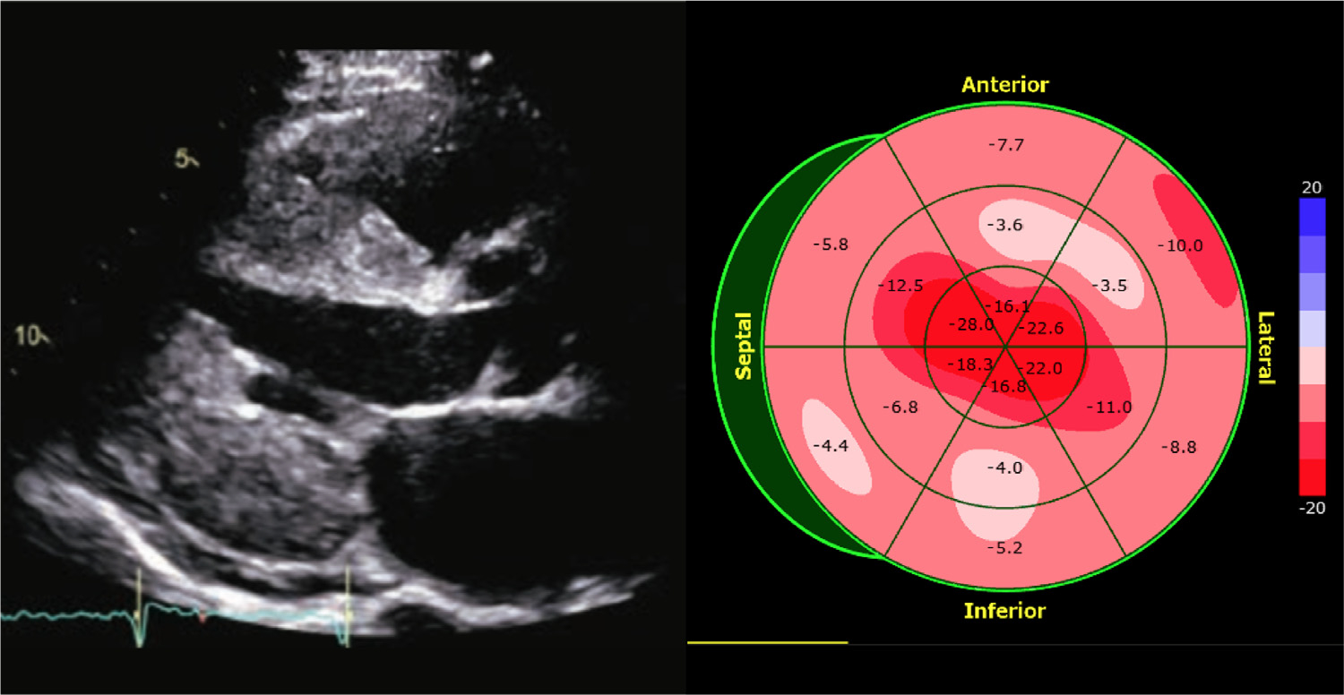
Example of an apical sparing pattern of LV strain on echocardiogram of a patient with suspected cardiac amyloidosis. (Left): parasternal long-axis view; (right): a bull’s eye display of longitudinal strain depicting preserved strain at the apex in the presence of severely reduced strain magnitude in mid- and basal LV segments.