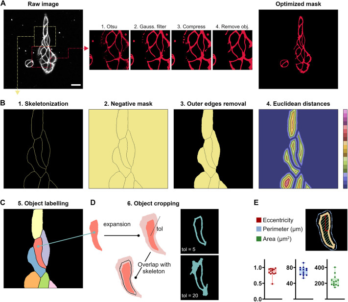
Fig. 2.
Working principles of image segmentation. (A) Optimization of the mask for the analysis of a parameter on cells. Starting from the raw image, different independent operations can be performed on the preliminary mask for optimization. (B,C) Working principle for the image segmentation. Steps 1 to 5 show the operations performed on the optimized mask to obtain a final image of individually labelled objects. (D) Consecutive steps for the cropping of individual objects. Depending on the tolerance parameter (tol) the user can expand or reduce the cropped object. For the plots, the box represents the 25–75th percentiles, and the median is indicated. The whiskers show the range. Each data point corresponds to an individual cell in Fig. 2A. (E) Object-specific morphological parameters calculated by the software. Scale bar: 5 µm.

