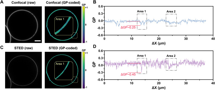
Fig. 7.
VISION can profile membrane fluidity with super resolution. (A,B) An example of a phase-separated GUV stained with Di-4-AN(F)EPPTEA imaged with confocal microscopy (A) and its corresponding profiled trajectory (B). (C,D) An example of phase-separated GUV imaged with STED microscopy (C) and its corresponding profiled trajectory (D). Scale bar: 3 µm distance. Images of GUVs reused from Sezgin et al. (2017) where they were published under a CC-BY 4.0 license.

