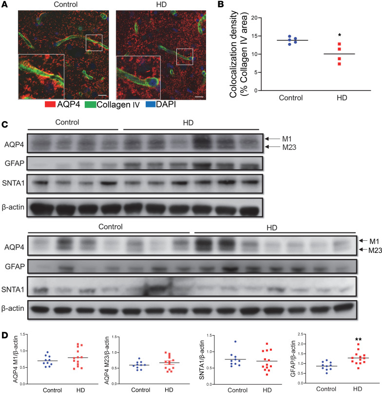
Figure 4. Reduced perivascular AQP4 localization accompanying astrogliosis in the human HD brain.
(A) Representative images of coimmunofluorescence staining of AQP4 (red) and collagen IV (green) in the caudate putamen of a patient with HD and age-matched control. Scale bar = 100 μm. Insets in A are 3 times enlarged from the original images. (B) Quantification of colocalized pixels of AQP4 (red in A) and collagen IV (green in A) in the caudate putamen of HD patients (n = 6) and age-matched controls (n = 4). *P < 0.05 vs. control by standard Student’s t test. (C) Western blots of AQP4, SNTA1, and GFAP in the human caudate samples from 13 HD brains and 10 control brains. (D) Quantification of AQP4 (both isoforms), SNTA1, and GFAP protein levels (ratio to the loading control β-actin) in the caudate samples. **P < 0.01 vs. control by standard Student’s t test.

