. 2024 Oct 2;16(10):e70723. doi: 10.7759/cureus.70723

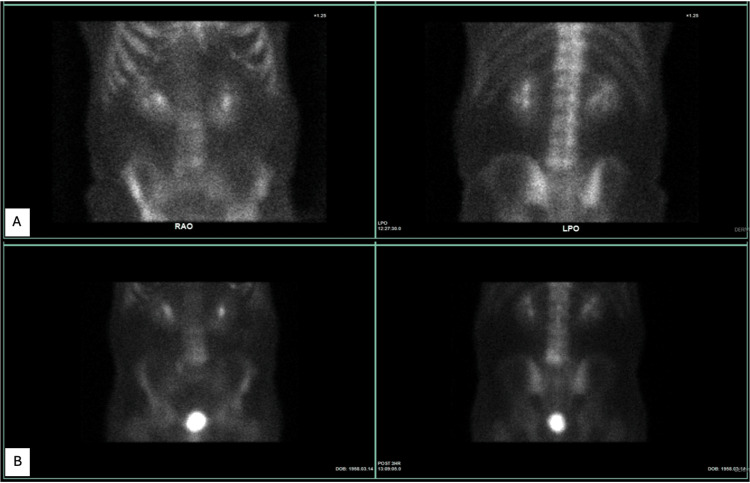
Figure 3. Technetium pyrophosphate nuclear scintigraphy was performed as part of the patient's work-up for the lesion. (A) The first scan performed 32 months after cancer diagnosis, which demonstrated mildly increased uptake in the left side of L5, corresponding to the lesion found on computed tomography in Figure 2. (B) Due to the rather large interval increase in the size of the L5 lesion and the appearance of a new lesion in L4, a repeat scan was performed 47 months after cancer diagnosis, which demonstrated a stable appearance of the mildly increased uptake in L5 identified previously, slightly increased uptake adjacent to this in the inferior margin of L4, and likely non-bridging diffuse idiopathic skeletal hyperostosis in the thoracic spine.

Figure 3