. 2024 Oct 2;16(10):e70723. doi: 10.7759/cureus.70723

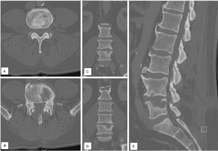
Figure 6. Repeat non-contrast computed tomography of the lumbar spine was performed 47 months after cancer diagnosis and demonstrated a marked interval expansion of the erosive L5 lesion with poorly defined margins, which now measured 30.4×22.6×13.8 mm. In addition, a hypodense, erosive lesion in the left side of L4 was also identified, which measured 4×13.9×4.3 mm. Figure 6A demonstrates an axial cut at the level of the L4 vertebral body lesion, while Figure 6B demonstrates the lesion in the L5 vertebral body. Figures 6C and 6D demonstrate coronal cuts, with Figure 6C showing both lesions and demonstrating a more anterior cut than the image presented in Figure 6D. Figure 6E demonstrates the L4 and L5 vertebral body lesions on a sagittal image.

Figure 6