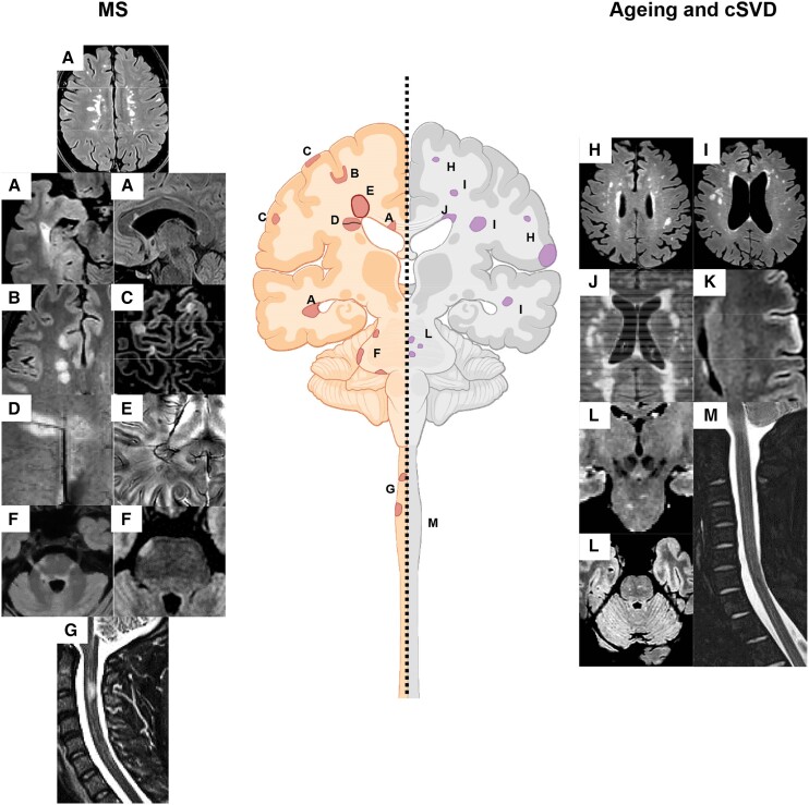
Figure 2.
Summary of the typical lesional MRI findings in multiple sclerosis compared to ageing and cerebral small vessel disease. Typical multiple sclerosis (MS) lesions include (A) periventricular lesions, (B) juxtacortical and cortical lesions, (C) white matter (WM) lesions showing the central vein sign (CVS), (E) paramagnetic rim lesion (PRLs), (F) infratentorial lesions mainly located at the periphery, close to the CSF, and (G) spinal cord lesions. Typical lesions occurring with ageing and cerebral small vessel disease (cSVD) include (H) subcortical WM lesions, (I) deep WM lesions, (J) periventricular lesions and ‘capping’, (K) cortical microinfarcts, (L) central pontine lesions and (L) no spinal cord lesions. See text for further details.

