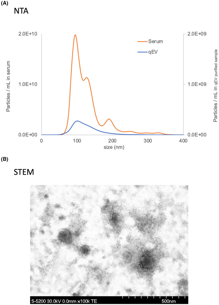
FIGURE 2.

(A) The EVs in PDAC serum (orange line) and purified EVs in PDAC serum by size exclusion chromatography (qEV column) (blue line) were analyzed using NTA. (B) The purified EVs were observed using STEM. EV, extracellular vesicle; NTA, nanoparticle tracking analysis; PDAC, pancreatic ductal adenocarcinoma; qEV, ; STEM, scanning transmission electron microscopy.
