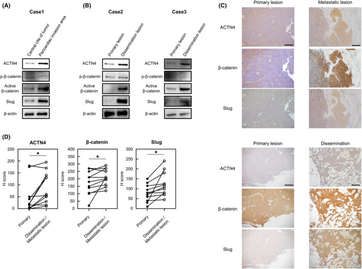
FIGURE 7.

Evaluation of ACTN4, β‐catenin, and Slug expression levels using surgical specimens. (A) Western blotting analysis of ACTN4, β‐catenin, and Slug expression between the pericardial invasion site and the non‐invasion site of a tumor from a thymoma type B3 patient. (B) Differential expression of ACTN4, β‐catenin, and Slug as indicated by western blotting between disseminated and primary lesions of a tumor from a thymoma type B2 patient. (C, D) Comparison of ACTN4, β‐catenin, and Slug expression by immunohistochemical staining between primary lesions and disseminated or distant metastatic lesions using surgical specimens (n = 11). Staining intensity was evaluated. Scale bars indicate 200 μm. *p < 0.05.
