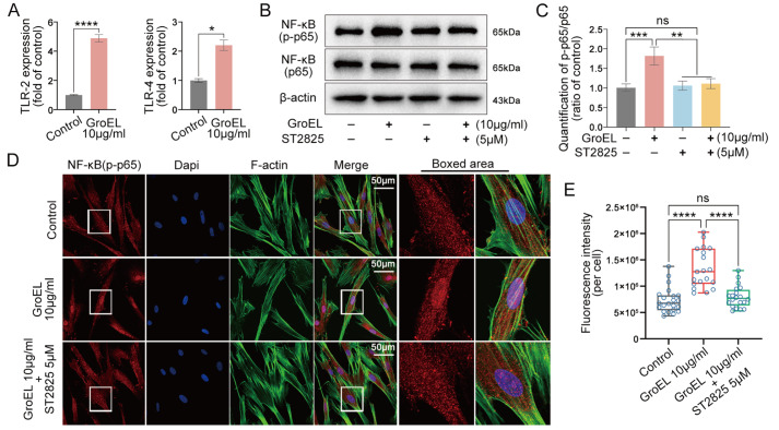
Figure 4 .
Inhibition of TLRs interferes with GroEL-induced NF-κB signaling
(A) qPCR showing the mRNA expressions of TLR-2 and TLR-4 in hPDLSCs induced by GroEL for 2 h. The data were derived from three separate experiments ( n=3). * P<0.05, **** P<0.0001. (B) Western blot images showing the protein expressions of NF-κB (p-p65) and NF-κB (p65) in hPDLSCs induced by GroEL after pretreatment with ST2825. The images were chosen from three separate experiments ( n=3). (C) Quantitative analysis confirming the protein changes in (B). Relative protein expression was normalized to that of β-actin. The data were derived from three separate experiments ( n=3). ** P<0.01, *** P<0.001. (D) Immunofluorescence images showing that ST2825 (5 μM) pretreatment attenuated GroEL-induced activation of NF-κB (p-p65) in hPDLSCs. The images were derived from three separate experiments ( n=3). (E) Quantitative analysis of total fluorescence density (OD/cell area) confirming the changes in the NF-κB (p-p65) protein in (D). The data were analyzed based on 19 cells from three separate experiments ( n=3). **** P<0.0001.

