
Fig. 5 ∣. Cobll1-deficient mice are leaner and display metabolically dysfunctional phenotypes.
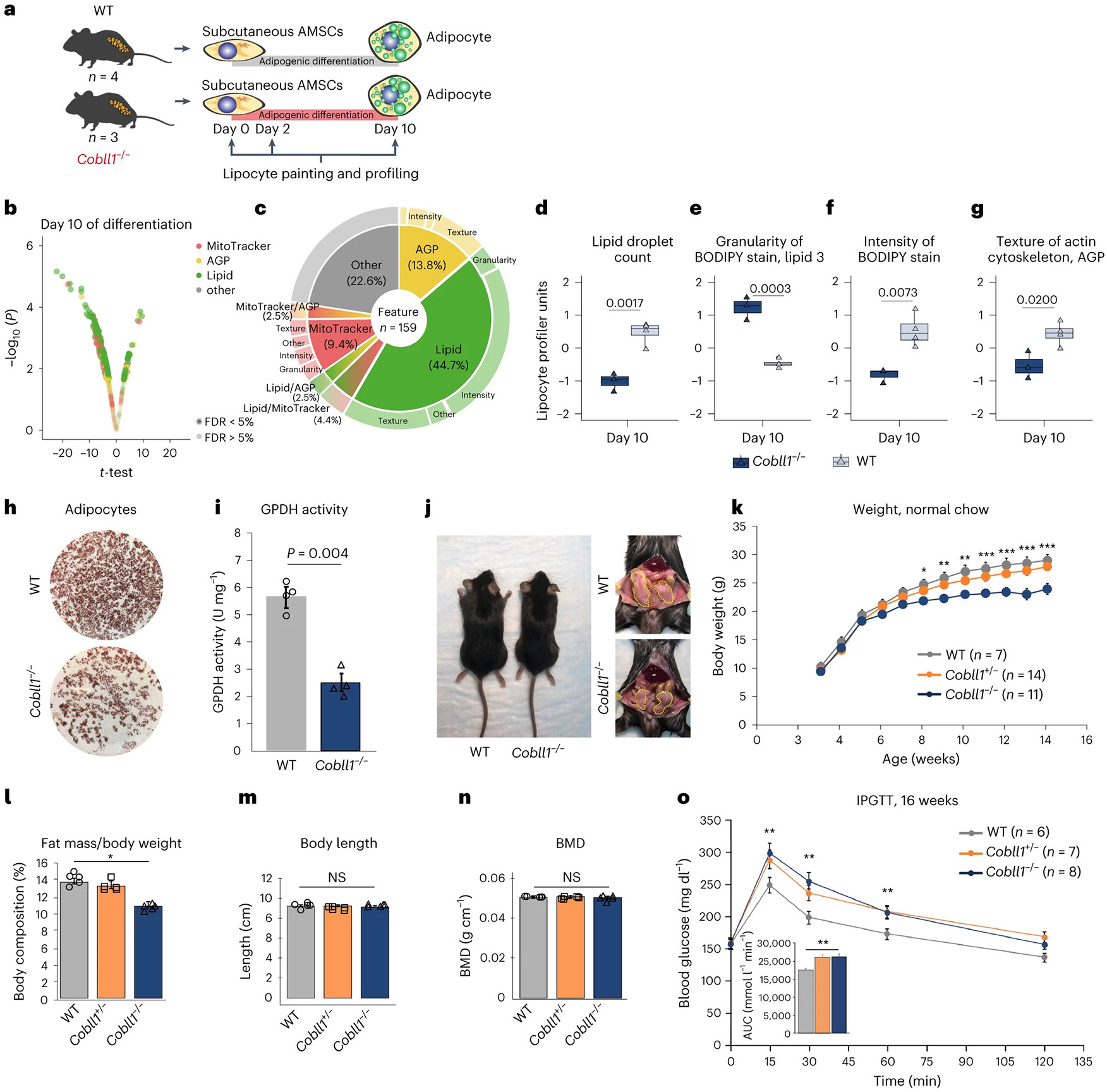
a, Schematic of differentiation and LipocyteProfiling at three time points (days 0, 2 and 10) of AMSCs derived from Cobll1−/− mice (n = 3) and WT mice (n = 4). b, Morphological profiles of the AMSCs of Cobll1−/− mice compared to the AMSCs of WT mice at day 10; two-sided t-test, significance level FDR ≤ 5%. c, Pie chart illustrating non-redundant differential features per channel and class of measurement comparing the AMSCs of Cobll1−/− and WT mice at day 10 of differentiation. d–g, Lipid droplet count (Cells_Children_LargeLipidObjects_Count) (d), granularity of BODIPY stain (Cells_Granularity_3_BODIPY) (e), intensity of BODIPY stain (Cells_Intensity_IntegratedIntensity_Lipid) (f) and texture of actin cytoskeleton (Cytoplasm_Texture_Entropy_AGP) (g) at day 10 of differentiation. Two-sided t-test; data represent the median ± 95% CI. h, Oil Red O staining of differentiated murine AMSCs. i, GPDH activity of differentiated murine AMSCs was assessed by measuring the decrease in NADH at 340 nm. Data represent the mean ± s.e.m. *P < 0.05 compared to the WT group. j, Representative photograph of 14-week-old WT and Cobll1−/− mice fed a normal chow. The yellow dotted lines delineate perigonadal WAT. k, Mouse body weight across time. Data are expressed as the mean ± s.e.m. *P < 0.05, **P < 0.01 and ***P < 0.001 compared to the WT group. l, Body composition (fat mass/body weight). Data are expressed as the mean ± s.e.m. *P < 0.05 compared to the WT group. m, Body length measurements of WT and Cobll1−/− mice (n = 6). Data are expressed as the mean ± s.e.m. *P < 0.05 compared to the WT group. NS, not significant. n, BMD analysed by dual energy X-ray absorptiometry (DEXA). Data are expressed as the mean ± s.e.m. *P < 0.05 compared to the WT group. o, IPGTT in WT and Cobll1−/− and Cobll1+/− mice. The inset graph shows the area under the curve (AUC) of the blood glucose concentration levels measured during IPGTT. Data represent the mean ± s.e.m. **P < 0.01 compared to the WT group. In i,k–o statistical significance was determined by Student’s t-test. In l–o the experiment was repeated independently three times with similar results.