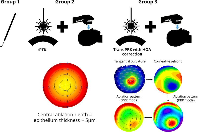
Figure 1.
Flowchart of the surgical procedures in each group. Left: Utilization of a reusable hockey knife. Center: Ablation pattern demonstrated by the central ablation and calculated by epithelium thickness plus 5 µm (yellow area). In the periphery, the ablation depth is higher as the laser light cannot enter at a perpendicular angle to the corneal surface. The lights therefore travel a longer way through the epithelium and the epithelium thickness is measured perpendicular to the surface of the cornea. Right: From corneal topography or tomography, a corneal wavefront is calculated, then imported into the laser planning software. Ablation pattern is assessed in PRK mode, if ablation depth in the cone (central red area) does not exceed 50 µm. A peripheral ablation of more than 50 µm was allowed as corneal thickness is usually thicker in this area.

