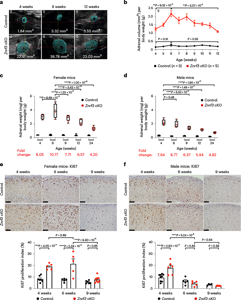
Fig. 1 |. Following an initial phase of significant hyperplasia, ZNRF3-deficient adrenal glands regress over time.
a, Ultrasound imaging provides a non-invasive method for measuring adrenal size in real time. Representative three-dimensional ultrasound images from a female control and a Znrf3-cKO mouse are shown at 4 weeks (initiation of study), 6 weeks (maximum volume) and 12 weeks (end of study) of age. b, Weekly ultrasound monitoring in female control and Znrf3-cKO mice identifies a phenotypic switch from hyperplasia to regression at 6 weeks of age. Statistical analysis was performed using one-way ANOVA followed by Tukey’s post hoc test. Error bars represent mean ± s.e.m. c, Adrenal weight measurements over time confirm significant adrenal regression in female Znrf3-cKO mice beginning after 6 weeks of age. d, In males, adrenal weight peaks earlier than females at 4 weeks and progressively declines with increased age. c,d, Each dot represents an individual animal. Box-and-whisker plots indicate the median (line) within the upper (75%) and lower (25%) quartiles, and whiskers represent the range. e, Proliferation as measured by Ki67 is significantly increased in adrenals of 4- and 6-week-old female Znrf3-cKO mice compared to controls. At the onset of adrenal regression (9 weeks), proliferation is significantly reduced in ZNRF3-deficient adrenals. f, Male Znrf3-cKO mice similarly exhibit hyperproliferation at 4 weeks of age. However, proliferation returns to baseline by 6 weeks, which is earlier than in females. e,f, Each dot represents an individual animal. Error bars represent mean ± s.e.m. Statistical analysis was performed using two-way ANOVA followed by Tukey’s multiple-comparison test. Scale bars, 100 μm.

