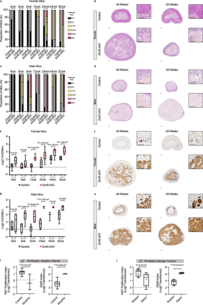
Extended Data Fig. 7 |. Immune cell recruitment in the adrenal gland is sex- and age-dependent.
(a, b) Histological evaluation of female control and Znrf3 cKO adrenal tissue based on H&E. Female Znrf3 cKOs continue to accumulate histiocytes with advanced aging at 44- and 52-weeks of age. (c-d) Male Znrf3 cKOs sustain high levels of histiocytes previously observed as early as 24-weeks of age. Quantification was performed using QuPath digital analysis based on the proportion of histiocyte area normalized to total adrenal cortex area. Scale bars, 100 μm. Data from 4- to 24-weeks of age was previously shown in Fig. 3f-i, and is included as reference. (e-f) In situ validation of myeloid cell accumulation based on IHC for CD68 in control and Znrf3 cKO adrenal tissue from female and (g-h) male cohorts at 44- and 52-weeks of age. Data from 4- to 24-weeks of age was previously shown in Fig. 4e-h, and is included as reference. Quantification was performed using QuPath digital analysis based on the number of positive cells normalized to total nuclei. Each dot represents an individual animal. Box and whisker plots indicate the median (line) within the upper (75%) and lower (25%) quartiles, and whiskers represent the range. Statistical analysis was performed on log2 transformed data using two-way ANOVA followed by Tukey’s multiple comparison’s test. Scale bars, 100 μm. (i) At 78-weeks of age, atrophic Znrf3 cKO adrenal glands have a significantly lower Ki67-index and higher CD68-index compared to age-matched controls. (j) In 78-week-old benign Znrf3 cKO adrenals, nodules have a significantly higher Ki67-index and lower CD68-index compared to the background gland. Each dot represents an individual animal. Box and whisker plots represent mean with variance across quartiles. Statistical analysis was performed using two-tailed Student’s t-test.

