. 2024 Nov 4;13:RP98458. doi: 10.7554/eLife.98458

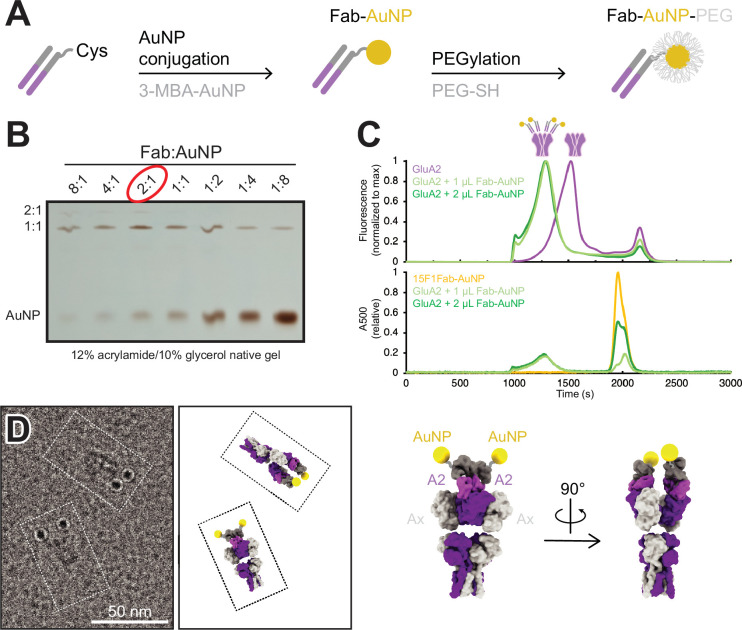
Figure 1. Development and characterization of anti-GluA2 Fab conjugated to gold nanoparticle (AuNP).

(A) Conjugation strategy for covalent linkage of AuNP to anti-GluA2 Fab. (B) Test-scale conjugations of Fab and AuNP at various Fab:AuNP ratios, run on native 12% acrylamide/10% glycerol gel. (C) Normalized fluorescence-detection size-exclusion chromatography (FSEC) traces of GFP-tagged GluA2 mixed with 1–2 µL of Fab-AuNP conjugate, measured at an excitation/emission of 480/510 nm (top; corresponding to GFP-tagged GluA2) or an absorbance at 500 nm (bottom; corresponding to AuNP). (D) Snapshot from single particle cryo-electron microscopy micrograph image showing two individual Fab-AuNP bound native mouse hippocampus AMPARs next to models depicting possible orientational views seen in image (PDB: 7LDD) (left). Model of anti-GluA2 Fab-AuNP bound to AMPAR with GluA2 in the B and D positions (right).

Figure 1—source data 1. PDF file containing original gels for Figure 1B, indicating relevant bands.
Figure 1—source data 2. Original files for gel analysis are displayed in Figure 1B.

Figure 1.

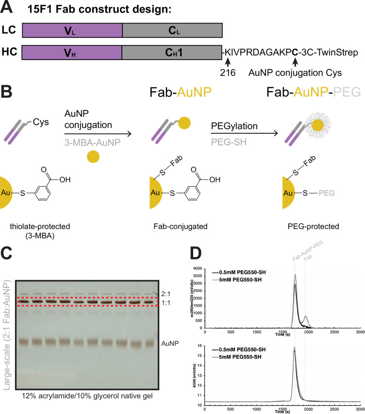
Figure 1—figure supplement 1. Preparation of PEGylated AuNP-15F1 Fab conjugate.

Figure 1—figure supplement 1.

(A) Design of anti-GluA2 15F1 Fab construct for gold nanoparticle (AuNP) conjugation. The light chain (LC) is included without modification, while the heavy chain contains an extension of the constant domain 1 to include a single hinge cysteine for AuNP conjugation, followed by a 3 C protease cleavage site and Twin-Strep tag. (B) Strategy for covalent conjugation and subsequent PEGylation of anti-GluA2 15F1 Fab and 3-MBA-protected AuNPs. (C) Full-scale conjugation of 15F1 Fab and AuNP using 2:1 Fab:AuNP ratio, with entire reaction run on native 12% acrylamide/10% glycerol gels (representative shown). The gel bands cut out for subsequent purification are denoted by the dashed red box. (D) Assessment of AuNP PEGylation with 0.5 or 5 mM PEG550-SH by size-exclusion chromatography (SEC), using HPLC measurement of tryptophan fluorescence (top) and AuNP absorbance at 500 nm (bottom).
Figure 1—figure supplement 1—source data 1. PDF file containing original gels for Figure 1—figure supplement 1C, indicating the relevant bands.
Figure 1—figure supplement 1—source data 2. Original files for gel analysis are displayed in Figure 1—figure supplement 1C.
Figure 1—figure supplement 2. Single particle cryo-electron microscopy (cryo-EM) of Fab-gold nanoparticle (AuNP) bound to native mouse hippocampal AMPAR.
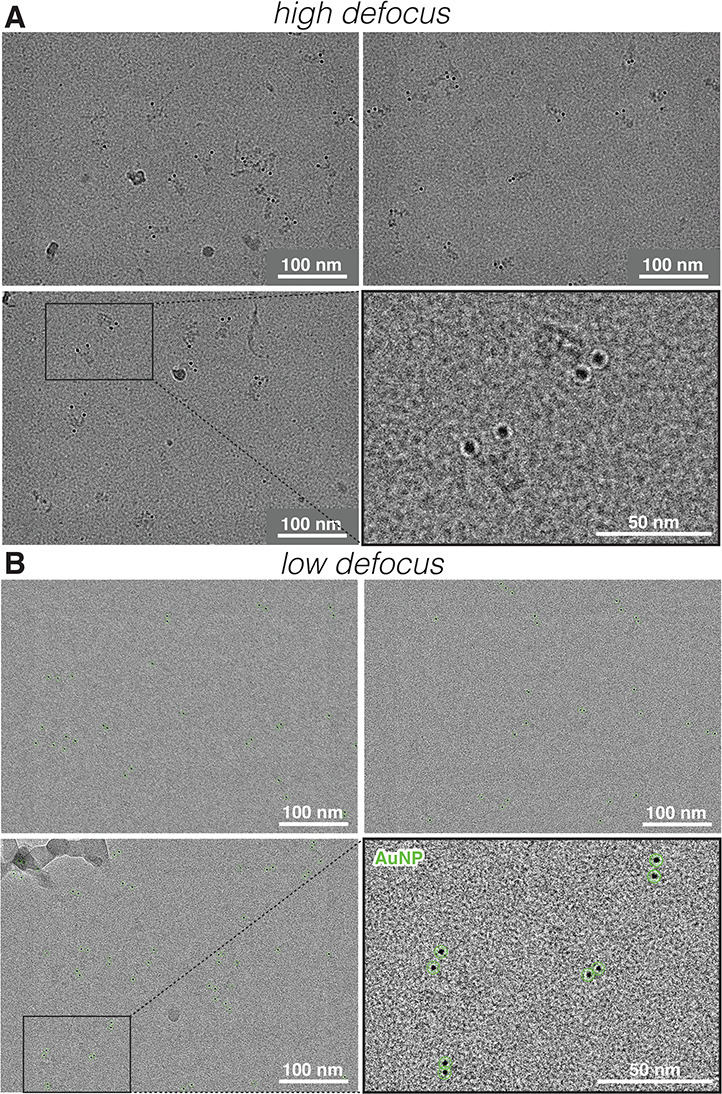
Figure 1—figure supplement 2.

Example single particle cryo-EM micrographs of native mouse hippocampal AMPAR bound to anti-GluA2 15F1 Fab-AuNP taken at (A) ‘higher’ (−4.5 to –5.0 µm) and (B) ‘lower’ (−1.0 to –1.2 µm) defocus ranges. Example automated bead-picking hits from low-defocus micrographs denoted with green circles.