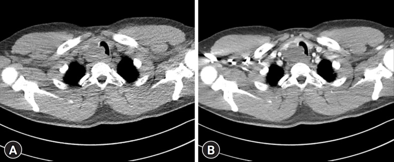
Fig. 2.
Neck computed tomography image reveals a lobulated low-density mass approximately 5 cm in size in the inferior portion of the right thyroid gland, extending to the upper mediastinum, and direct invasion of the upper trachea, causing luminal narrowing. No enhancement is visible in the mass (A) before and (B) after contrast administration.

