Figure 1.

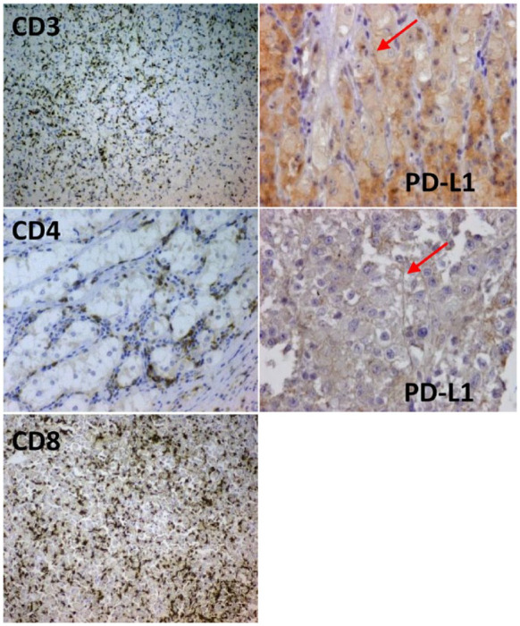
Left: lymphocytic infiltration. Right: two examples with membrane-positive staining for PD-L1.
PD-L1, programmed death-1 ligand.

Left: lymphocytic infiltration. Right: two examples with membrane-positive staining for PD-L1.
PD-L1, programmed death-1 ligand.