
Figure 1. The ion permeation path of A2iQ/γ2(KKEE).
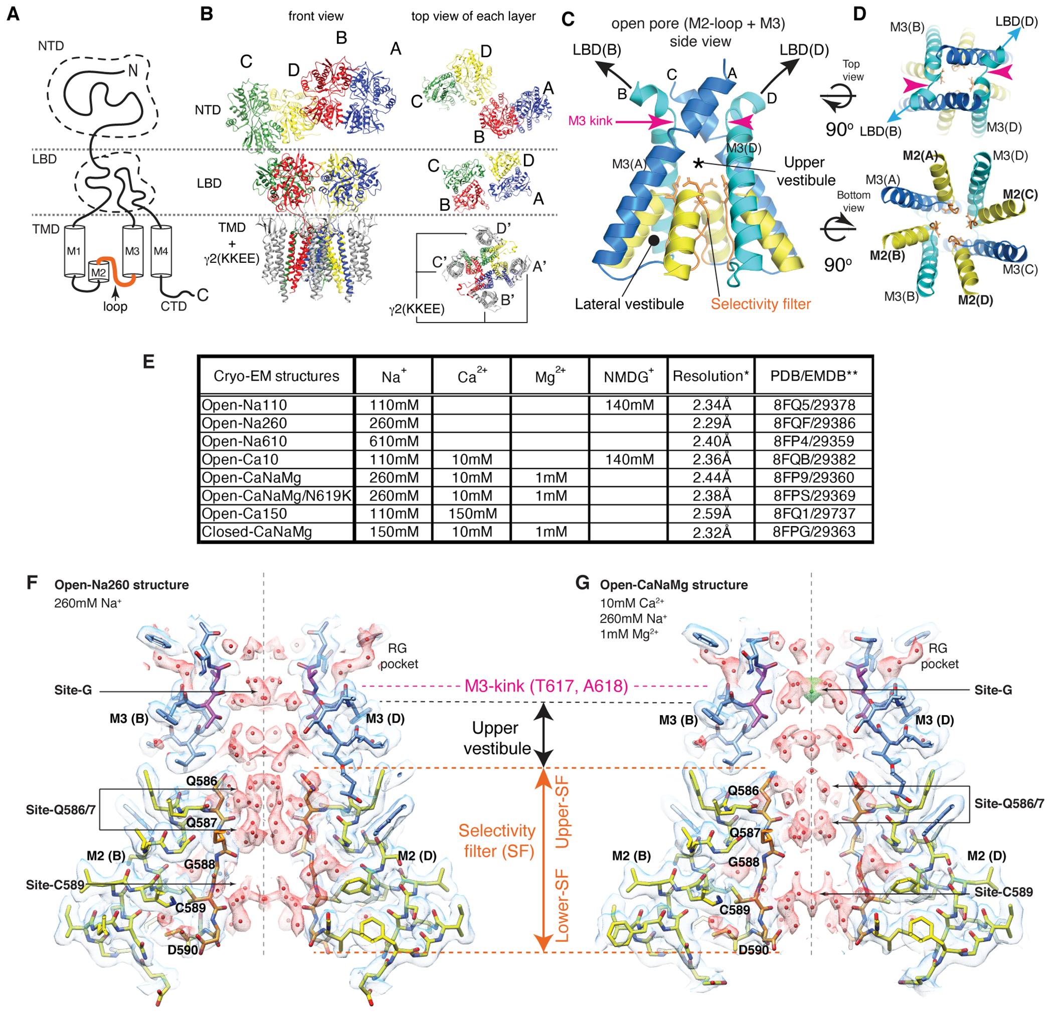
A. Domain organization of AMPAR subunit. The orange loop contains the selectivity filter. B. Tetrameric assembly of A2iQ/γ2(KKEE), whose global structure is indistinguishable from the WT complex. GluA2 subunits; A=blue, B=red, C=green, and D=yellow. Four γ2(KKEE)s are in gray and their positions are labeled A’, B’, C’ and D’. C. Architecture of the open pore. Blue=M3 of A/C subunits, cyan=M3 of B/D subunits, yellow=M2, and orange=selectivity filter. D. Top: The open pore viewed from the extracellular space. Bottom: The selectivity filter viewed from the cytoplasmic side. E. Summary of ion conditions used in each cryo-EM structure. (*) C2 map resolutions of the transmembrane region are listed. See Table 1 and Supplementary Table 1 for details of refinement statistics. (** ) PDB/EMDB codes are for the transmembrane region. F and G. The central slices of the pore of Open-Na260 (E, map threshold=6.0σ) and Open-CaNaMg (F, map threshold=3.3σ). The cryo-EM density map (mesh) and the atomic model are superimposed. Putative water and Ca2+ are in red and green spheres, respectively. Regions of intense density accumulation are indicated as sites-G, -Q586/587, and -C589. Dashed gray lines are pore symmetry axes. The RG pocket indicates a cavity filled with water that is on the rear side of site-G and commonly found in the open structures.