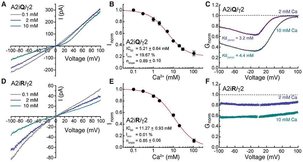
Figure3. External Ca2+ block of GluA2/γ2 and its voltage dependence.

A and D. Current records observed in presence of 10mM glutamate and 100μM CTZ at a voltage range of −100 to +100 mV in presence of 0.1mM (gray), 2mM (light blue) and 10mM (dark blue) Ca2+ for A2iQ/γ2 (Patch # 220914p7) and A2iR/γ2 (Patch # 221025p2) receptors. B and E. Inhibition plots of block by external Ca2+ of A2iQ/γ2 (B, n=8) and A2iR/γ2 (E, n=6) receptors at −100 mV. Data are presented as mean values ± SEM. C and F. Conductance-voltage plots of block by 2 mM or 10 mM external Ca2+ of A2iQ/γ2 (C) and A2iR/γ2 (F) receptors. The voltage-dependence of block of A2iQ/γ2 receptors was well fit by a single permeant ion blocker model (red line) whereas block of A2iR/γ2 receptors was voltage-insensitive.
