
Figure 2.
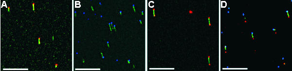
Combed DNA detection with QD. DNA molecules were modified with biotin or digoxigenin at one or both ends, stained with YOYO-1 (green) and combed. Biotin was detected with streptavidin QD 565 (D) or 605 (A and C) (shown in red) and digoxigenin was detected with mouse anti-digoxigenin and anti-mouse QD 655 (shown in blue). Four different labeling schemes were performed: biotin at one end (A), digoxigenin at one end (B), biotin at both ends (C), biotin at one end and digoxigenin at the other end (D). The QD images were processed from a 60 s long image movie (60 images), each pixel intensity is the maximum value of this pixel in the image stack. DNA image and QD maximum were then overlaid. The horizontal bar represents 10 μm.