Figure 5.
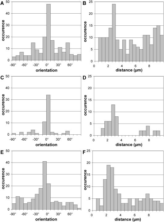
Preferential layout of QD pairs. DNA molecules were modified with biotin at both ends (A–D) or biotin at one end and digoxigenin at the other end (E and F) and combed. Biotin was detected with streptavidin QD 565 or 605 and digoxigenin was detected with mouse anti-digoxigenin and anti-mouse QD 655, respectively. QD images were analyzed using a program written in MATLAB. The angle between a QD pair and the combing direction (A, C and E) and the distance between both QD of the pair (B, D and F) were measured. Results are reported for QD 605 pairs (A, B, C and D) and for pairs involving one QD 565 and one QD 655 (E and F). In C and E, only QD pairs for which the distance is between 1.5 and 4.0 μm are represented. In D and F, only QD pairs for which the angle is between −10° and +10° are represented.

