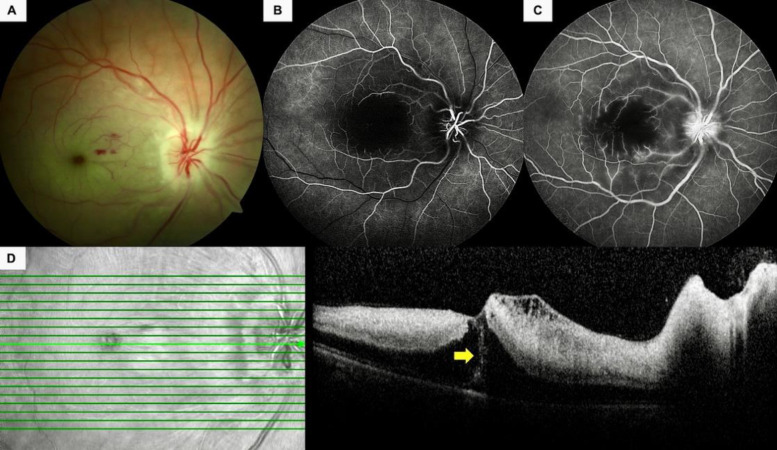
Figure 3.
A 34-year-old pregnant woman (Case 9) (Table 2) presented with painless, sudden-onset vision loss in her right eye. (A) Color fundus photographs depicted an infarcted macula, pale and swollen optic nerve head, and a few intraretinal hemorrhages. (B) The early phase of a fluorescein angiogram revealed the nonperfused foveola and late filling of the arterioles. (C) Some pinpoint leakages from the macular retinal arterioles and faint optic disc leakage was noted in the late-phase fluorescein angiogram. (D) Spectral-domain optical coherence tomography revealed retinal thickening, increased hyperreflectivity of the inner retinal layers, and an intraretinal hyperreflective line (yellow arrow).

