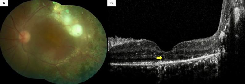
Figure 4.
A 10-year-old boy (Case 26) (Table 2) with Coats’ disease displayed (A) extensive retinal exudation and photocoagulation scars in his left eye, along with a serous retinal detachment at the temporal macular aspect. Furthermore, the color fundus photograph was blurred because of a posterior subcapsular cataract. However, (B) an intraretinal hyperreflective line was visible (yellow arrow) on the spectral-domain optical coherence tomographic section passing through the fovea, along with a massive exudation at the temporal aspect.

