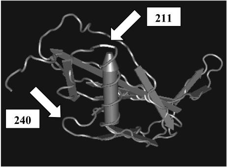
FIG. 3.
β-Domain of streptokinase from S. dysgalactiae subsp. equisimilis (49), showing a single α-helix (tube-shaped arrow) and β-folded sheets consisting of multiple strands (flat gray arrows). White arrows indicate the localization of the codons homologous to codons 211 and 240 in the S. uberis PauA sequence.

