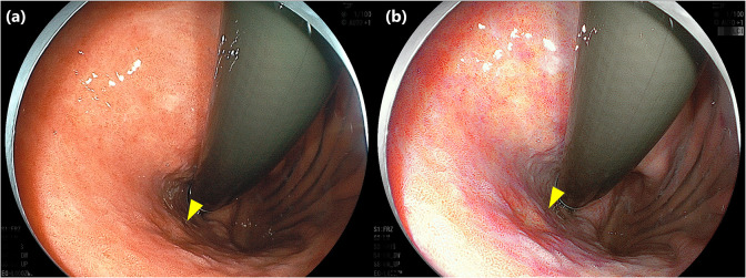
Fig 4. Representative images of early gastric cancer in which the visibility was classified as poor visibility on WLI but improved to good visibility on LCI.
Type 0-Ⅱc early gastric cancer, 9 mm in size, located on posterior wall of the upper gastric body (yellow triangle). The mean visibility score on WLI was 1.7, which was classified as poor visibility (a), but the mean visibility score on LCI improved to 3.0 (b). WLI, White light imaging; LCI, Linked color imaging.

