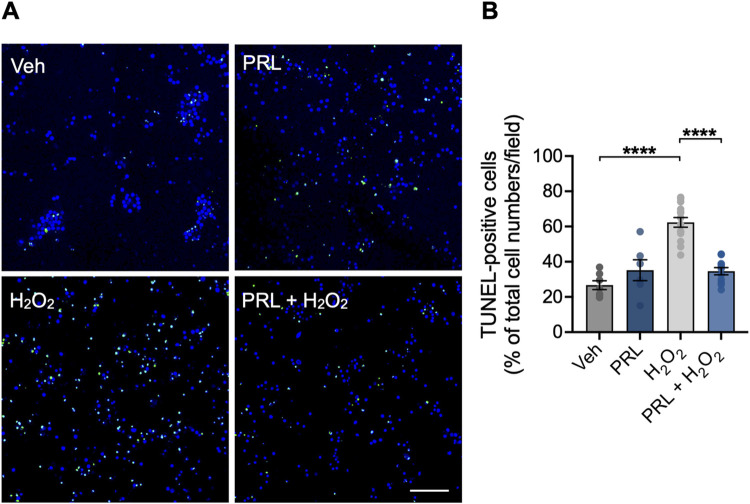
Fig 5. Effect of PRL on H2O2-induced apoptosis in mouse hippocampal neurons.
Hippocampal neurons were pre-incubated for 24 h with 100 nM prolactin (PRL) or vehicle, followed by treatment with 100 μM H2O2 or vehicle for 24 h. Cells were stained for apoptosis using the TUNEL assay (green), and nuclei were conterstained with DAPI (blue). (A) Representative images of TUNEL staining in hippocampal neuronal cultures treated with vehicle, PRL, and H2O2. Scale bar 200 μm. (B) Quantification of TUNEL-positive cells. Bar plot shows the percentage of neurons positive to TUNEL staining per image. (n = 3). ****p<0.0001 vs vehicle or indicated group.

