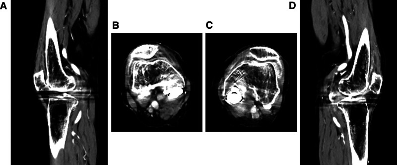
Figure 4.
(A) The sagittal images of contrast-enhanced CT for the right knee joint after steroid administration. (B) The axial images of contrast-enhanced CT for the right knee joint after steroid administration. (C) The axial images of contrast-enhanced CT for the left knee joint after steroid administration. (D) The sagittal images of contrast-enhanced CT for the left knee joint after steroid administration.

