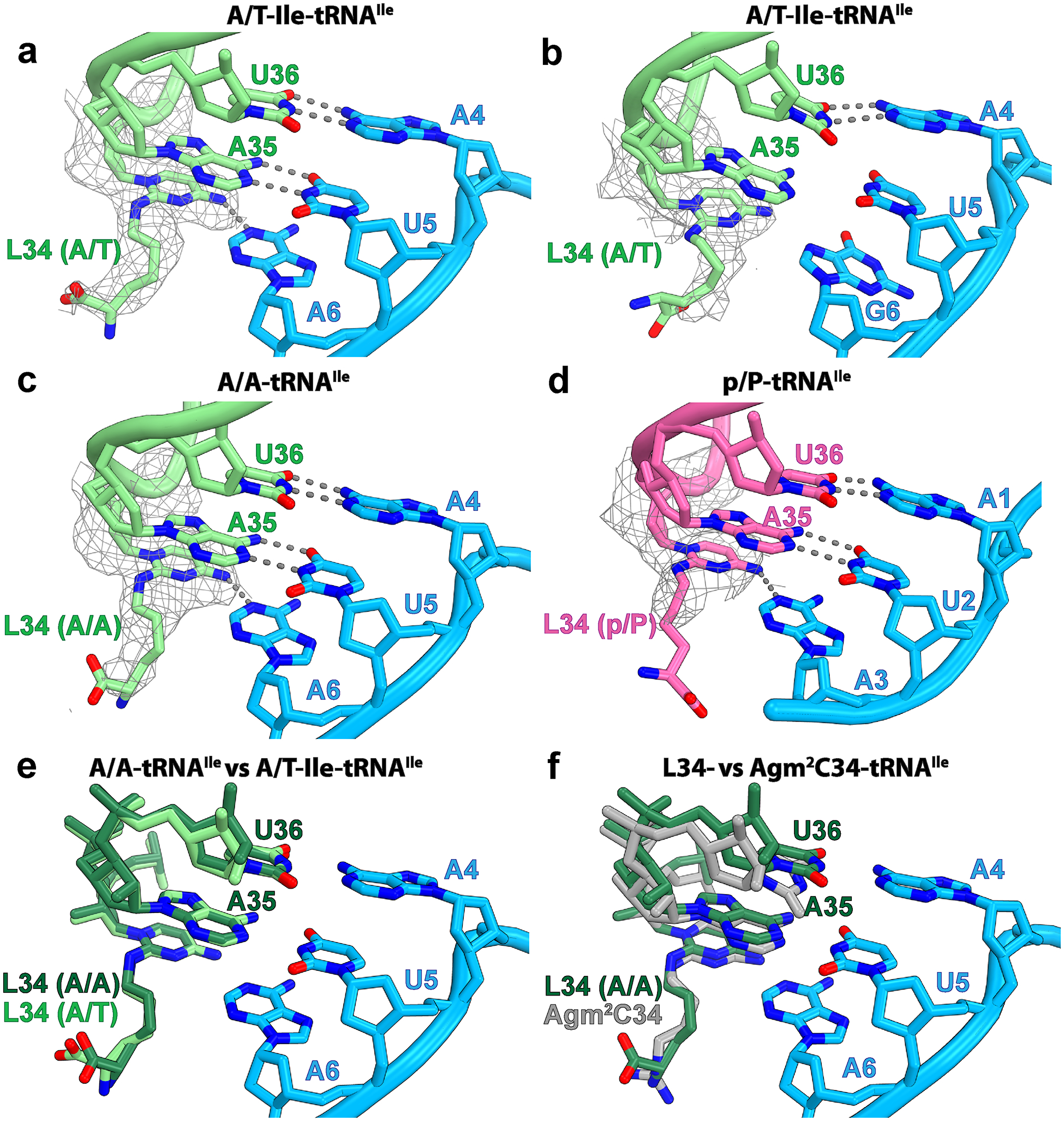
Fig. 2: Role of lysidine 34 in tRNAIleLAU during decoding.

a, Base pairs between the A/T-Ile-tRNAIleLAU (light green) and the AUA codon (light blue) in structure I. b, Same as in a with the AUG codon in structure II. c, Base pairs between the accommodated A-site tRNAIleLAU and the AUA codon in structure III. d, Base pairs between the P-site tRNAIleLAU (magenta) and the AUA codon in structure IV. The Coulomb potential density of lysidine 34 (L34), contoured at 2.9σ, is shown as gray mesh. The gray dashed lines show putative hydrogen bonds. e, Relative conformation of the anticodon loop, including L34, of tRNAIleLAU in the A/T (light green, structure I) and A/A (dark green, structure III) bound states. f, Structure alignment of A-site tRNA2Ile containing agm34 from Haloarcula marismortui (gray, PDB 4V8N (ref.36)) with the A-site lysidine-containing tRNAIleLAU from E. coli (dark green, structure III).
