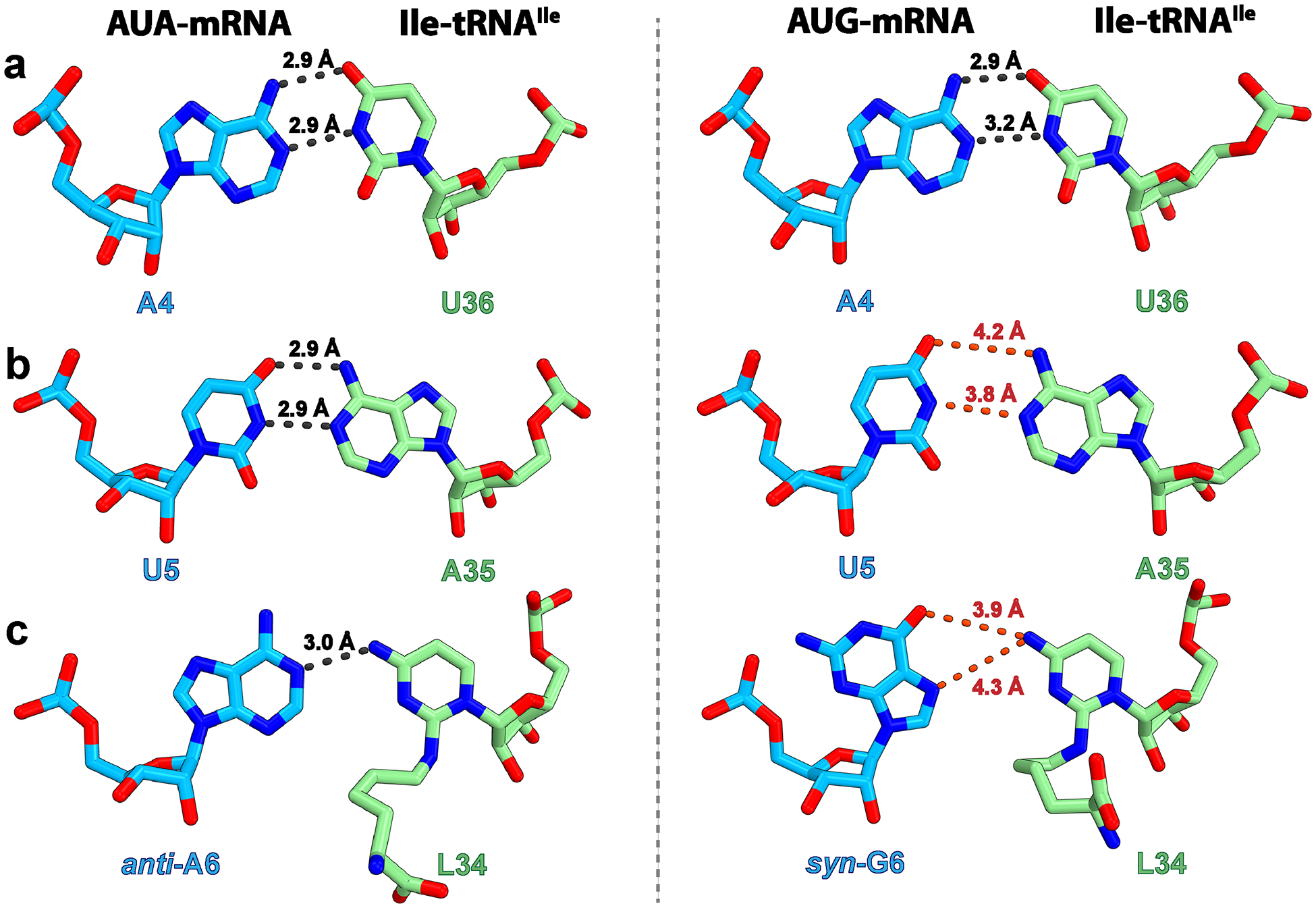
Fig. 3: Conformation of codon–anticodon base pairs with the cognate AUA and near-cognate AUG codons.

a–c, Base pair at the first (a), second (b) or third (c) position of the codon formed between the E. coli Ile-tRNAIleLAU and the cognate AUA (left, structure I) or near-cognate AUG (right, structure II) codon. The gray dashed lines indicate distances that are conducive to hydrogen bond formation (<3.5 Å threshold42), whereas those above this threshold, and therefore unlikely to form, are red.
