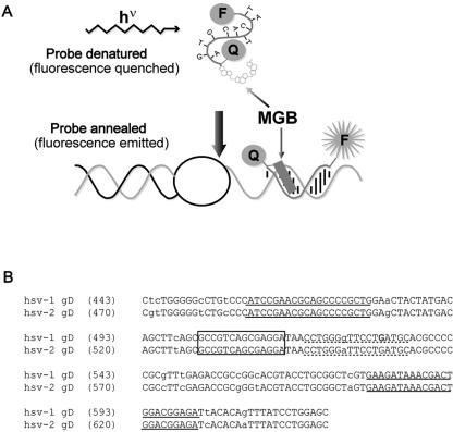
FIG. 1.
HSV Eclipse probe design. (a) Schematic diagram of an Eclipse hybridization probe. MGB indicates the minor groove-binding moiety. F indicates the fluorescent dye, and Q represents the quenching molecule. (b) Alignment of HSV-1 (gi:330064) and HSV-2 (gi:517467) glycoprotein D (gD) sequences surrounding the amplified fragment. Lowercase letters indicate polymorphisms between HSV-1 and -2. Primer sequences are underlined with solid lines. The dashed underline indicates the original probe sequence. Within this sequence, the bold G indicates the novel polymorphism that was identified. The final probe sequence is boxed.

