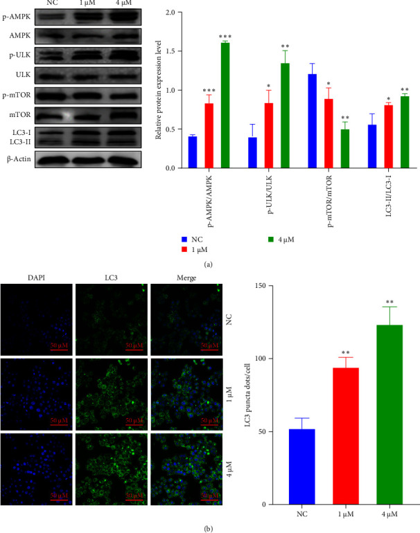
Figure 4.

The inhibitory effect of shikonin on EC9706 cell autophagy. (A) Western blot analysis was used to determine the protein (p-AMPK, p-ULK, p-mTOR, and LC3) levels in EC9706 cells. ∗P < 0.05, ∗∗P < 0.01, and ∗∗∗P < 0.001 versus the normal control (NC) (n = 3). (B) The LC3 puncta were examined using confocal microscopy and were quantified. LC3 is shown in green. 4′,6-diamidino-2-phenylindole (DAPI) is shown in blue, which stained the nuclei. Confocal microscope was taken at ×20.
