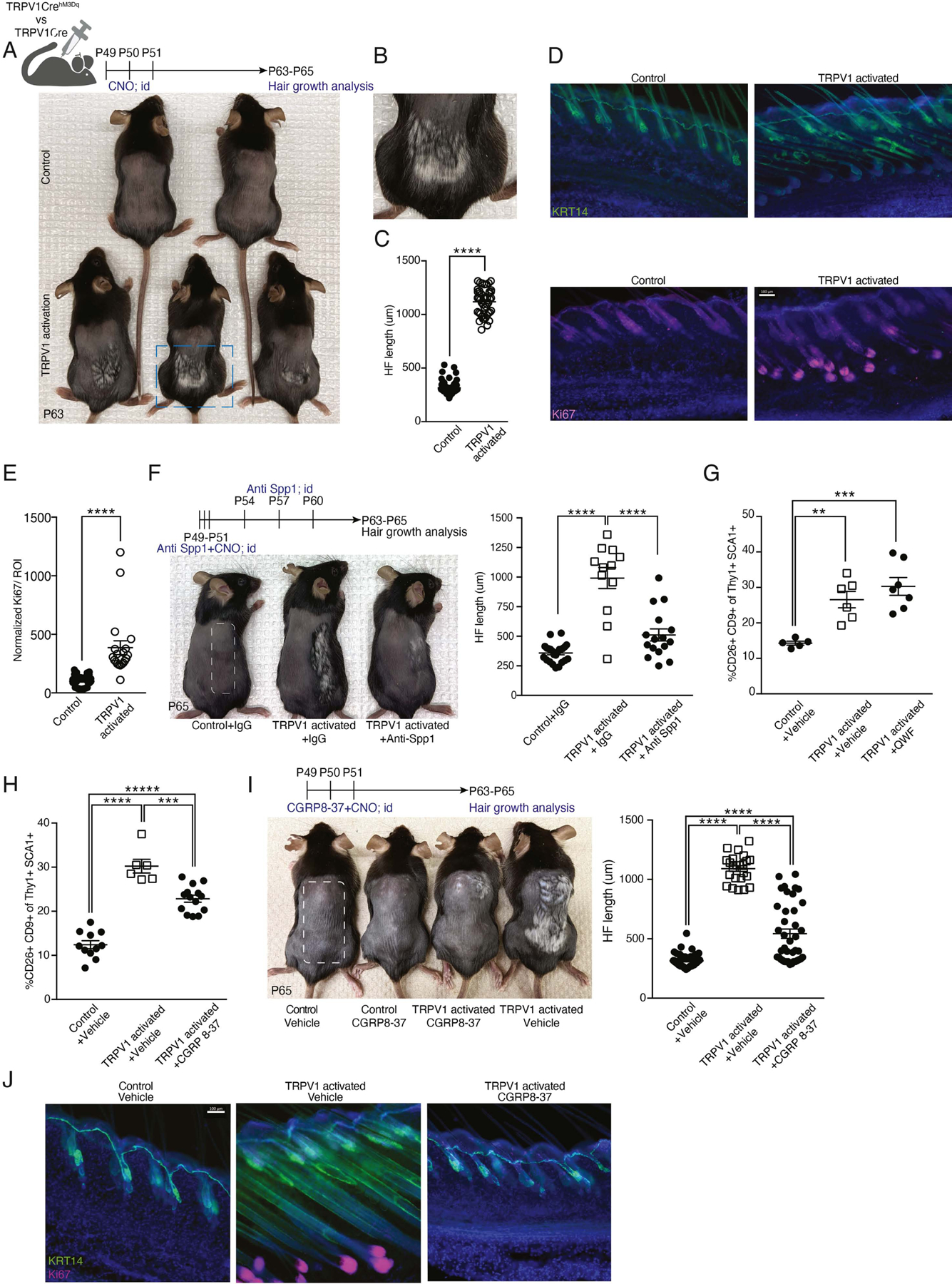
Figure 2: Repeated cutaneous TRPV1 activation triggered a CGRP-dependent acceleration of hair growth.

(A) Diagram of experimental design and representative images from shaved TRPV1 activated mice and controls. Mice monitored for hair coat growth for 12 days after the last TRPV1 activation with CNO (postnatal day 63).
(B) Enlarged dashed rectangle from panel A depicting hair growth in a TRPV1 activated mouse.
(C) HF length quantification of dorsal skin from mice treated as in A. Data combined from 6 to 8 HF per mouse, taken from 6 mice per group (****p<0.0001).
(D) Representative immunofluorescent images of dorsal skin from mice treated as in A. Staining with anti-keratin14 (KRT14; green) marking epidermal layer and Ki67 (purple) marking the HF bulb. DAPI stain in blue. Scale bar 100μm.
(E) Quantification of normalized Ki67 MFI signal in the peri-follicular area of dorsal skin from mice treated as in A. Data combined from 4 regions of interest (ROI) per mouse taken from 5 TRPV1 activated mice and 9 controls (****p<0.0001).
(F) Diagram of experimental design and representative images from TRPV1 activated mice and controls treated with CNO on half of their back skin (marked with dashed rectangle). Anti-Spp1 neutralizing antibody or IgG control were injected intradermally to the same skin area. Data are combined from 4 HFs per mouse collected from 6 controls, 3 TRPV1 activated + IgG treated and 4 TRPV1 activated + anti-Spp1 treated mice (****p<0.0001).
(G-H) Percentage of CD9+CD26+ dermal fibroblasts from TRPV1 activated mice and their controls intradermally injected with (G) QWF (SubP antagonist) or vehicle (**p=0.005, ***p=0.0004), (H) CGRP8–37 (CGRP antagonist) or vehicle (***p=0.0001, ****p<0.0001).
(I) Diagram of experimental design and representative images from TRPV1 activated mice and controls pre-treated intradermally with CGRP8–37 or vehicle and monitored for hair coat growth for 14 days. Quantification of HF length from 6 to 8 HFs per mouse, taken from 3–6 mice per group (****p<0.0001).
(J) Representative immunofluorescent images of dorsal skin from mice treated as in I. Stained with anti-KRT14 (green), Ki67 (pink), and DAPI (blue). Scale bar, 100μm.
