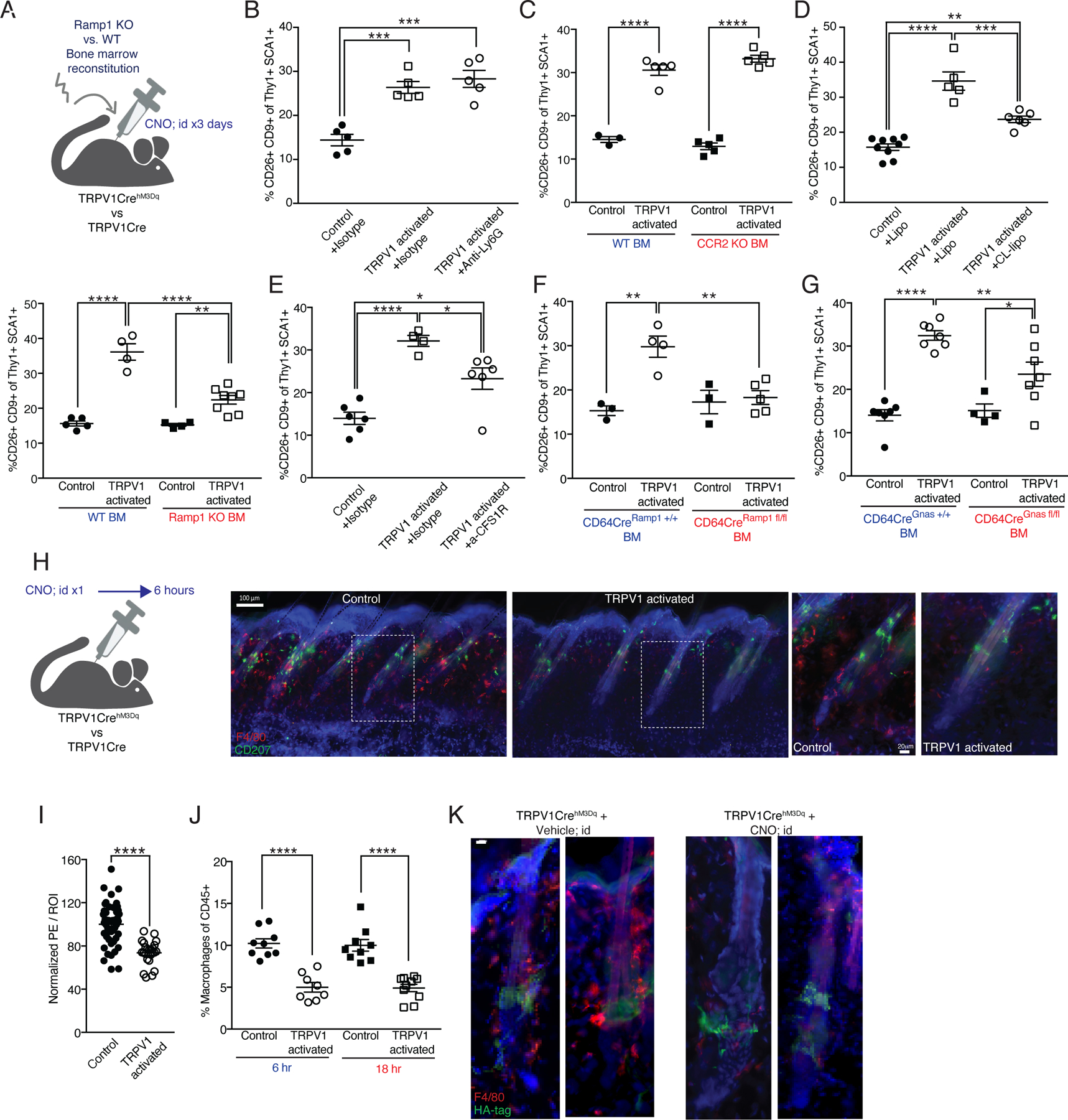
Figure 3: Macrophages rapidly respond to cutaneous TRPV1 activation and partake in mediating its effects on dermal fibroblasts.

(A-G) Quantification of CD9+CD26+ dermal fibroblasts from TRPV1 activated mice and controls given three daily intradermal CNO injections.
(A) (Upper) Diagram of experimental design. (Lower) Mice reconstituted with Ramp1 KO or wild type BM (**p=0.005,****p<0.0001).
(B) Mice pretreated systemically with isotype control or anti-Ly6G antibody (***p=0.0005).
(C) Mice reconstituted with CCR2 heterozygous or homozygous KO BM (****p<0.0001).
(D) Mice pretreated systemically with clodronate (CL) or control liposomes (lipo) (**p=0.002,***p=0.0004,****p<0.0001).
(E) Mice pretreated systemically with anti-CSF1R or isotype control (*p=0.029,****p<0.0001).
(F) Mice reconstituted with CD64CreRamp1+/+ (control) or CD64CreRamp1fl/fl BM (**p=0.003, 0.002).
(G) Mice reconstituted with CD64CreGnas+/+ (control) or CD64CreGnasfl/fl BM (*p=0.033,**p=0.007,****p<0.0001).
(H) Diagram of experimental design and representative immunofluorescent images of dorsal skin from TRPV1 activated mice and controls after a single intradermal CNO injection. 150μm sections stained with anti-F4/80 (red), anti-CD207 (Langerin, green), and DAPI (blue). Scale bar 100μm. Dashed rectangles indicate enlarged area in images on the right 20μm scale bar.
(I) Quantification of F4/80 normalized MFI signal in the peri-follicular area. Data combined from 8 region of interest (ROI) per mouse taken from 5 control and 3 TRPV1 activated mice treated as in H (****p<0.0001).
(J) Percentage of dermal macrophages from TRPV1 activated and control mice at 6 and 18 hours after a single intradermal CNO injection (****p<0.0001).
(K) Representative immunofluorescent images of HF from mice treated as in H. 50μm sections stained with anti-HA-tag (green), F4/80 (red), and DAPI (blue). Scale bar 10μm.
