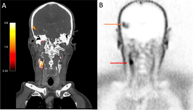
Fig. 2.
18F-GP1 Positron Emission Tomography images of a patient with an acute ischaemic stroke caused by large artery atherosclerosis. Images from an 81-year old gentleman presenting with headache and confusion. Panel (A) shows fused computed tomography and positron emission tomography images with PET uptake seen as red/orange. Corresponding PET only images in Panel (B). Both panels show18F-GP1 uptake in right carotid artery (Panel (A)—red/orange colour and Panel (B)—red arrow) indicating acute plaque rupture with thrombus embolization causing right middle cerebral artery infarct (Panel (A)—red/orange colour, Panel (B)—orange arrow).

