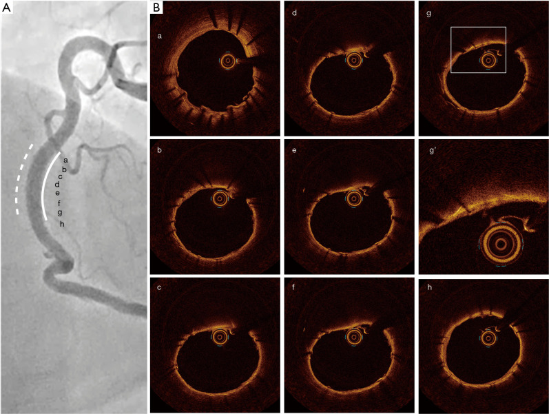
Figure 2.
Follow-up coronary angiography and OCT imaging at 7 months after PCI. (A) In-stent restenosis did not occur at segment receiving covered stent (white line) [a-h correspond to OCT images in (B)]. Dotted white line indicates the implanted drug-eluting stent. (B) Protruding of CN was not observed at segment receiving covered stent by OCT imaging (g’ is an enlargement of the frame of g). OCT, optical coherence tomography; PCI, percutaneous coronary intervention; CN, calcified nodule.

