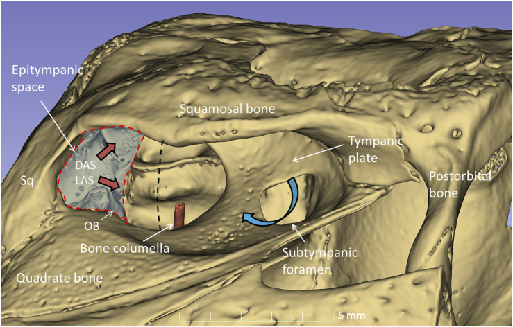
FIGURE 10.
µCT-3D rendering of the right bony external ear in a seven-month-old C. rhombifer - lateral view. There are two foramina and communication pathways between the meatal chamber and middle ear: one larger that houses the columellar shaft (red) and one smaller one, the subtympanic foramen, that is located anteriorly and aerates the anterior ear drum (blue arrow). A posterior epitympanic space (red interrupted line) contains muscles connected to the tympanic membrane and extracolumella (tensor and stapedius muscles) and the levator and depressor auricular superior muscles (LAS, DAS, red arrows). LAS and DAS muscles are attached to the acoustic plate. The superior earflap is attached to the lateral boundaries of the squamosal and postorbital bones. The dashed dark line indicates the margin between anterior and posterior portions of the tympanic membrane shown in Figure 11.

