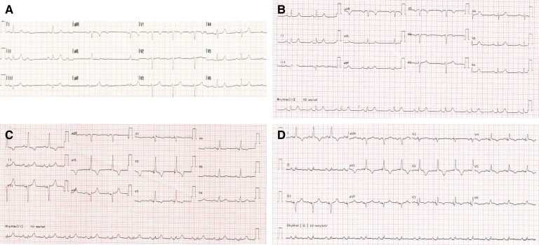
Figure 1.
Electrocardiograms (ECG)s at different time points. (A) Pre-hospital ECG showed intermittent 2:1 atrioventricular block. (B) ECG at admission showed sinus rhythm with prolonged PR interval (206 ms). (C) Day 0 post-procedure ECG showed selective left bundle branch pacing. (D) Day 3 post-procedure ECG showed selective left bundle branch pacing with mild ST elevation and T wave inversion in V2–6.

