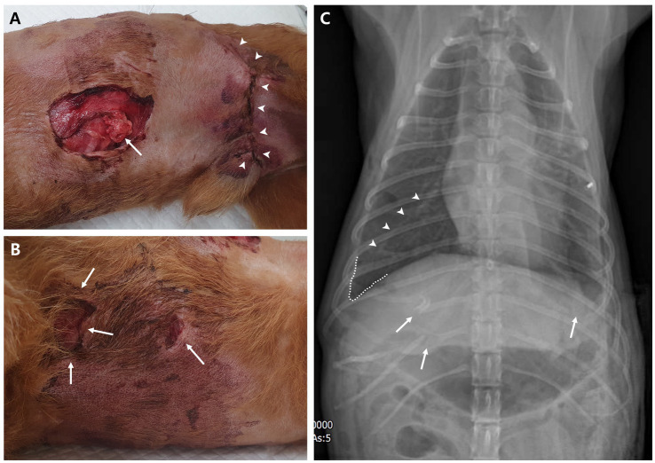
Figure 2.
Physical examination and diagnostic image at presentation. (A) Ventral left thoracic open wound with herniated soft tissue (arrow) and left inguinal wound (arrowheads), which had been closed at the local animal hospital; observed in the right lateral recumbent position. (B) Ventral right thoracic open wounds (arrows) near the sternum observed in the same position. (C) Dorsoventral thoracic radiography reveals fractures in the right 11th–12th rib and left 8th rib (arrows). A fissure line (arrowheads) between the right middle and caudal lobe of the lung and blunting of the right costophrenic sulcus indicate lung retraction due to pneumothorax or pleural effusion, such as hemothorax. In addition, overall lung opacity is elevated.

