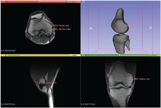
Figure 1.
Sample images in 3D Slicer showing the identification of the MCL insertion. Selected points were used to determine the tibial and femoral centroids, and the distance between the tibial and femoral centroids was calculated as the insertion and used when measuring the length of the MCL during the single-leg squat. MCL, medial collateral ligament; 3D, 3-dimensional; ant, anterior; post, posterior.

