
Fig. 2.
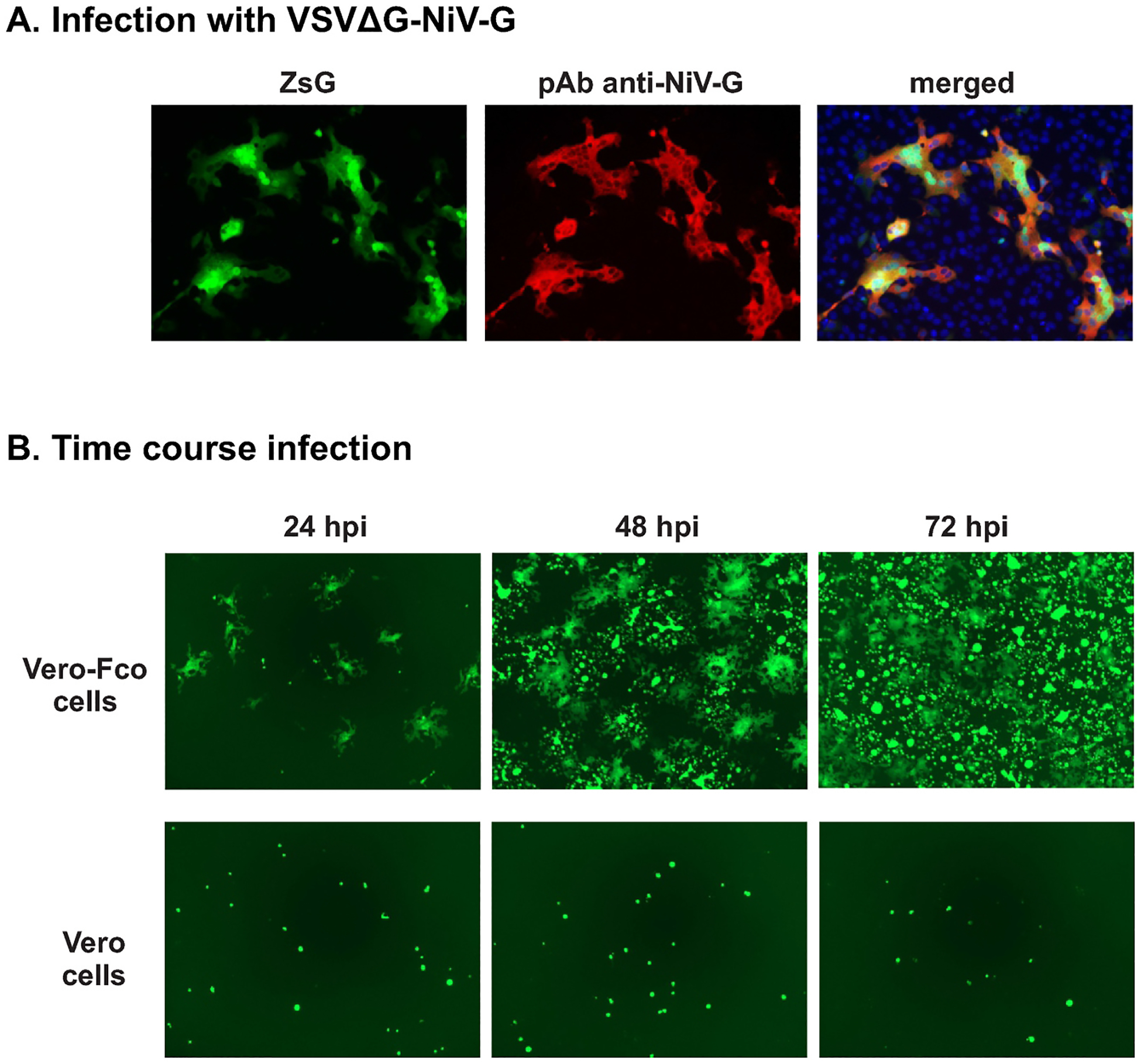
Infection of cells with rVSVΔG-NiV-G
A) rVSVΔG-NiV-G replication in Vero-Fco cells: Representative IFA images (magnification 10 ×; EVOS microscope) of Vero-Fco infected with rVSVΔG-NiV-G and probed with anti-NiV G antibody. B) Single-cycle replication in rVSVΔG-NiV-G in Vero-E6 cells: Vero-Fco and Vero-E6 cells were infected with rVSVΔG-NiV-G and ZsG fluorescence images (magnification 10 ×; EVOS microscope) were taken 24 h post infection (hpi), 48 hpi, and 72 hpi.