
Fig 4.
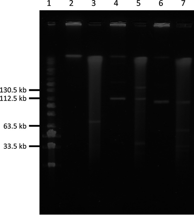
Confirmation of the presence of linear plasmids by S1 nuclease pulsed-field gel electrophoresis (PFGE) of optrA-positive enterococci. Lane 1, PFG MidRange Marker; lane 2, 2001206 (undigested); lane 3, 2001206 (digested); lane 4, 2001564 (undigested, ~114 kb linear plasmid present); lane 5, 2001564 (digested, ~114 kb linear plasmid present); lane 6, 2008300 (undigested, ~106 kb plasmid present); and lane 7, 2008300 (digested, ~106 kb plasmid present).