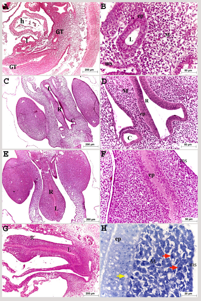
Fig. 1.
Colorectum of a quail embryo at day 3 (A, B) and 4 of incubation (C-H). Histomorphology. Longitudinal (A, B, G, H) and cross paraffin sections (C-F). H&E (A–G) and methylene blue stains (H). Images B, D, F and H were details of images A, C, E, G. Gut tube (GT), heart (h), liver (l), colorectum (R), caecum (C), lumen of colorectum (L), pseudostratified epithelium (ep), vacuoles (yellow arrow), mesenchyme (M), mitotic division (red arrow) and mesothelium (ms)

