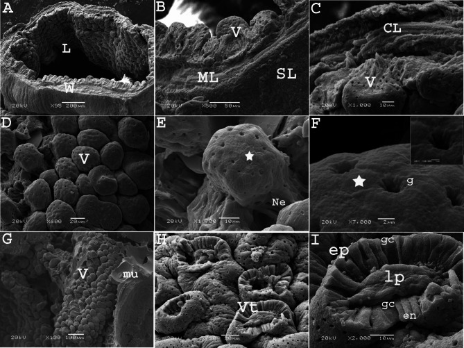
Fig. 10.
Scanning electron micrographs (SEM) of the colorectum of a quail embryo at day 17 of incubation. Images B, C, E, F, H and I were details of images A, D and G. The rectal wall (W), lumen (L), villi (V), muscular layer (ML), circular muscle layer (CL), serosa (SL), villi neck (Ne) goblet opening (g), honey-comb shaped arrangement of enterocytes (star), villi with losing tips (Vt), mucous secretion (mu), epithelium (ep), goblet cells (gc), lamina propria (lp), and enterocytes (en)

