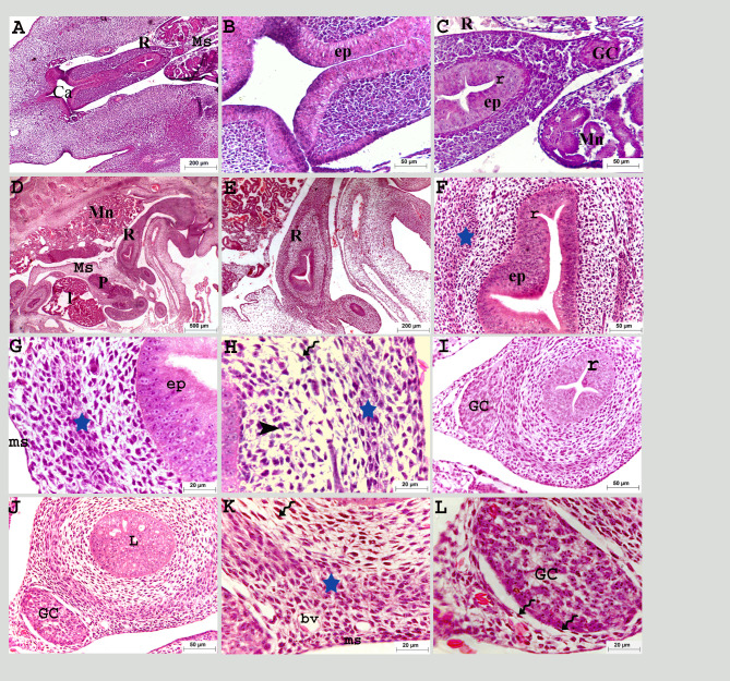
Fig. 2.
Colorectum of a quail embryo at day 5 (A-C) and 6 of incubation (D-L). Histomorphology. Cross (A-C, I-L) and longitudinal paraffin sections (D-F). H&E stain (A–I). Images B, C, E-I, K, and L were details of images A, D and J. Colorectum (R), cloaca (Ca), mesentery (Ms), pancreas (P), liver (l), mesonephros (Mn), pseudostratified epithelium (ep), previllous ridges (r), ganglionic cells (GC), mesenchymal condensation (star), telocytes (twisted arrows), mesenchymal cells (black arrow head) and mesothelium (ms), obliterated lumen (L), and blood vessels (bv)

