Fig. 3.
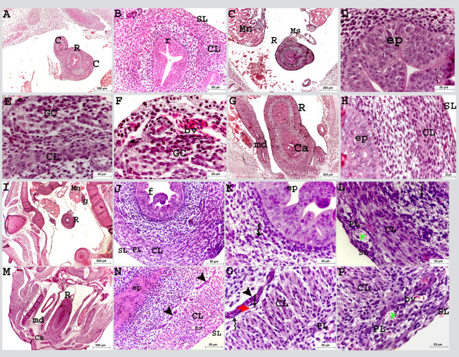
Colorectum of a quail embryo at day 8 (A-H) and 9 of incubation (I-P). Histomorphology. Longitudinal (A, B) and cross paraffin sections (C-P). H&E stain (A–P). Images B, D-F, H, J-L, K and N-P were details of images A, C, G, I and M. Colorectum (R), caecum (C), mesonephros (Mn), mesentery (Ms), previllous ridge (r), mesonephric duct (md), pseudostratified epithelium (ep), epithelium folds (f), circular muscular layer (CL), solitary groups of myoblasts (black arrow head), ganglionic cells (GC), cloaca (Ca), gonad (g), telocytes (twisted arrows), telopodes (red arrow head), myenteric plexus (PL) consisted of nerve cell bodies (green arrow head), blood vessel (bv), and serosa (SL)

