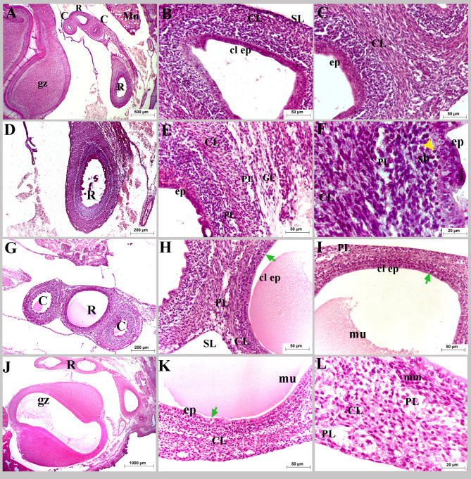
Fig. 4.
Colorectum of a quail embryo at day 10 (A-F) and 11 of incubation (G-L). Histomorphology. Longitudinal paraffin sections (A-L). H&E stain (A-L). Images B, C, E, F, H, I, K and L were details of images A, D, G and J. Colorectum (R), caeca (C), gizzard (gz), mesonephros (Mn), pseudostratified epithelium (ep), simple columnar epithelium (cl ep), enteroendocrine cell (green arrow), muscularis mucosa (mm), circular muscular layer (CL), ganglionic cells (GC), myenteric plexus (PL), mucous secretion (mu), and serosa (SL)

