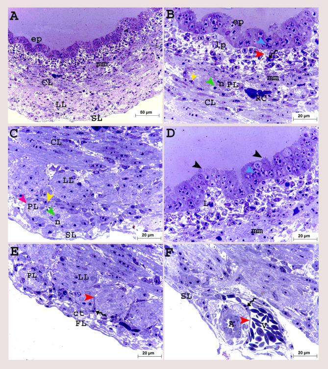
Fig. 5.
Colorectum of a quail embryo at day 12 of incubation (A-F). Histomorphology. Cross semithin sections (A-F). Toluidine blue stain (A-F). Images B-F were details of image A. Pseudostratified epithelium (ep), epithelium budding (black arrow head), lamina propria (lp), lymphocyte (L), mitotic divisions (blue arrow head), muscularis mucosa (mm), circular (CL), longitudinal (LL) muscular layers surrounded with telocytes (twisted arrows), telopodes (red arrow head), myenteric plexus (PL) consisted of capsule (purple arrow head) contained nerve cell bodies (n and green arrow head) flattened cells (yellow arrow head), artery (A), vein (V), red blood cell (RC), serosa (SL) consisted of connective tissue layer (ct) and layer of flattened cells (FL)

