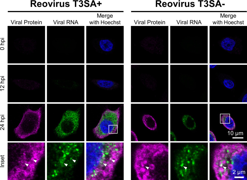
Fig 1.
HCR assay detects reovirus RNA at late stages of infection in cultured cells. HeLa cells were adsorbed with 100 PFU/cell of reovirus strains T3SA+ or T3SA− for 1 h. At the times post-adsorption shown, cells were fixed and stained for reovirus S3 RNA (green) by HCR and viral protein (magenta) by indirect immunofluorescence. Cells were counterstained with Hoechst dye (blue) and imaged using confocal microscopy. Representative images are shown. Arrowheads indicate sites of colocalization. Scale bar, 10 µm (single cell) or 2 µm (inset).

