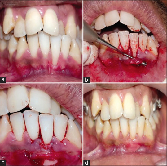
Figure 3.

(a) Case 2: Clinical photograph showing gingival recession in tooth number 31 (b) elevation of modified bridge flap (c) graft sutured at recipient site with additional horizontal adapting sutures (d) six months postoperative clinical photograph of recipient site of case 2
